Case report |
Peer reviewed |
Demonstration of porcine circovirus type 2 in the testes and accessory sex glands of a boar
Evidencia de circovirus porcino tipo 2 en los testículos y glándulas sexuales accesorias de un semental
Démonstration du circovirus porcin type 2 dans les testicules et les glandes sexuelles accessoires d'un verrat
T. Opriessnig, DVM; C. Kuster, DVM, PhD; P. G. Halbur, DVM, PhD
TO, PGH: Department of Veterinary Diagnostic and Production Animal Medicine, College of Veterinary Medicine, Iowa State University, Ames, Iowa. CK: Kuster Research and Consulting, Geneseo, Illinois. Corresponding author: Dr Patrick G. Halbur, Department of Veterinary Diagnostic and Production Animal Medicine, College of Veterinary Medicine, Iowa State University, Ames, IA 50011; Tel: 515-294-1137; Fax: 515-294-3564; E-mail: pghalbur@iastate.edu.
Cite as: Opriessnig T, Kuster C, Halbur PG. Demonstration of porcine circovirus type 2 in the testes and accessory sex glands of a boar. J Swine Health Prod. 2006;14(1):42-45.
Also available as a PDF.
SummaryPorcine circovirus type 2 (PCV2) antigen was demonstrated in lungs, lymphoid tissues, intestines, and testes and accessory sex glands of an 11-month-old boar from a boar stud with a history of recent illness and infertility. At necropsy, the lungs failed to collapse and there was moderate cranioventral bronchopneumonia. Microscopically, there was severe multifocal suppurative and histiocytic bronchointerstitial pneumonia with severe peribronchiolar lymphoid hyperplasia. Mycoplasma hyopneumoniae nucleic acids were demonstrated in lung by polymerase chain reaction (PCR), and moderate numbers of Pasteurella multocida type D were isolated from the lungs. There was mild to moderate lymphoid depletion and histiocytic replacement of follicles in lymphoid tissues associated with PCV2 antigen. Immunohistochemistry revealed PCV2-specific staining in the cytoplasm of macrophages and fibroblast-like cells in the interstitium of the seminal vesicles, and PCV2 nucleic acids were demonstrated in semen by nested PCR. These findings suggest that PCV2 was part of the disease complex that contributed to the illness in this boar. | ResumenEl antígeno del circovirus porcino tipo 2 (PCV2 por sus siglas en inglés) se detectó en los pulmones, tejido linfoide, intestinos, testículos y glándulas sexuales accesorias de un macho de 11 meses de edad de un centro de inseminación artificial que recientemente había reportado un episodio de enfermedad e infertilidad. A la necropsia los pulmones no colapsaron y se encontró una bronconeumonía cranioventral moderada. Microscópicamente, se detectó una neumonía severa bronco intersticial histiocítica supurativa y multifocal con hiperplasia linfoide peribronquiolar. El ácido nucleico del Mycoplasma hyopneumoniae se detectó en pulmón mediante la reacción en cadena de la polimerasa (PCR por sus siglas en inglés) y un número moderado de Pasteurella multocida tipo D se aisló de los pulmones. Se encontró depleción linfoide de leve a moderada y reemplazo histiocítico de los folículos en el tejido linfoide asociado al antígeno PCV2. La inmunohistoquímica reveló una tinción específica al PCV2 en el citoplasma de los macrófagos y células similares al fibroblasto en el intersticio de las vesículas seminales y el ácido nucleico del PCV2 se detectó en semen mediante PCR anidado. Estos hallazgos sugieren que el PCV2 fue parte del complejo de la enfermedad que contribuyó al padecimiento de este macho. | ResuméDe l'antigène de circovirus porcin type 2 (CVP2) a été démontré dans les poumons, tissus lymphoïdes, intestins, testicules et les glandes sexuelles accessoires d'un verrat de 11 mois provenant d'un centre d'insémination avec un historique de problème récent de maladie et d'infertilité. À l'autopsie, les poumons ne s'affaissaient pas normalement et présentaient une bronchopneumonie cranioventrale modérée. Microscopique-ment, on pouvait noter une pneumonie broncho-interstitielle suppurée multifocale et histiocytaire sérieuse de même qu'une hyperplasie lymphoïde péribronchiolaire importante. Des acides nucléiques de Mycoplasma hyopneumoniae ont été démontrés dans le poumon par amplification en chaîne par polymérase (PCR en anglais), et des nombres modérés de Pasteurella multocida type D ont été isolés des poumons. Il y avait une déplétion lymphoïde légère à modérée et un remplacement des follicules par des histiocytes dans les tissus lymphoïdes associés avec de l'antigène de CVP2. L'immunohistochimie a révélé une coloration spécifique au CVP2 dans le cytoplasme de macrophages et de cellules semblables à des fibroblastes dans l'interstitium des vésicules séminales, et des acides nucléiques de CVP2 ont été démontrés dans la semence par PCR nichée. Ces observations suggèrent que le CVP2 était impliqué en partie dans la maladie de ce verrat. |
Keywords: swine, boar,
fertility, porcine cirocvirus type 2, Mycoplasma hyopneumoniae, PCV2
Search the AASV web site
for pages with similar keywords.
Received: January
28, 2005
Accepted: March
11, 2005
Porcine circovirus type 2 (PCV2) in- fection is commonly associated with postweaning multisystemic wasting syndrome (PMWS), which primarily affects pigs 6 to 20 weeks of age.1,2 In mature pigs, PCV2 infection is commonly subclinical and considered to be of minor importance. The hallmark lesions associated with PCV2 infection are lymphoid depletion and histiocytic inflammation in lymphoid organs, with or without lymphohistiocytic inflammation in lung, liver, heart, intestine, kidney, or other organs.2
Porcine circovirus type 2 is considered a sporadic cause of reproductive failure, associated with abortions and increased incidence of stillborn and mummified fetuses.3-5 Severe, diffuse, nonsuppurative myocarditis, associated with large amounts of PCV2 antigen as demonstrated by immunohistochemistry (IHC) methods, is commonly observed in affected fetuses,3-5 whereas reports regarding PCV2-associated lesions in the dams are lacking. Little is known about the effect of PCV2 on boars or the significance of circulation of PCV2 in boar studs. Seven-month-old, experimentally infected boars shed PCV2 nucleic acids in semen for at least 47 days post infection, as determined by nested polymerase chain reaction (PCR).6 This report describes the diagnostic investigation of infertility in a boar stud and provides evidence that PCV2 may play a role in suboptimal fertility.
Case description
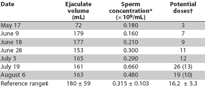
An 11-month-old boar was presented to the Veterinary Diagnostic Laboratory at Iowa State University (ISU-VDL; Ames, Iowa). The boar had a history of producing fewer sperm per collection than expected (Table 1), with the majority of ejaculates discarded due to poor motility as determined by phase contrast microscopy over the 12-week period prior to necropsy. While 19 to 26 doses of semen per collection would generally be considered within the normal range for a boar of this age,7 only the last two collections approached these levels. The previous five collections ranged from only three to 12 doses. The last two collections (July 19 and August 6) were preceded by at least 2 weeks of sexual rest. Had the boar been on a routine weekly collection schedule during the entire period, fewer doses per collection would have been expected on July 19 and August 6.
Table 1: Ejaculate characteristics of an 11-month-old boar during a 3-month period in a boar stud with a history of recent illness and infertility of boars
* Sperm concentration determined by photometry. † Prior to the July 19 and August 6 collections, the boar had 2 weeks sexual rest. The estimated weekly dose production is shown in parentheses. ‡ Cameron (1987).7 Mean +/- SD determined in 8- to 12-month-old boars (n = 15); values adjusted to reflect once-weekly collection schedule. |
At the time of presentation, the boar had a fever (body temperature 40.5 degrees C), rapid and labored breathing, and elevated heart rate persisting for 2 days. The boar originated from an isolated boar stud routinely tested for porcine reproductive and respiratory syndrome virus (PRRSV), pseudorabies virus, and Brucella species. Boars were obtained from multiple sources of varying herd health status. All boars were tested in isolation for PRRSV by enzyme-linked immunosorbent assay (ELISA) (HerdChek PRRS ELISA; Idexx Laboratories Inc, Westbrook, Maine). Those with positive titers were retested prior to entering the main stud and were allowed to enter only if the ELISA results were below the positive cutoff, ie, sample-to-positive (S:P) ratio < 0.4. For boars that were ELISA-positive in isolation or boars known to have been ELISA-positive prior to shipment to the boar stud, semen was tested for PRRSV by PCR8 to minimize the risk of having boars shedding PRRSV in their semen enter the main stud. Routine serology for Mycoplasma hyopneumoniae was not performed prior to entering the stud; however, some sources were known to be free of M hyopneumoniae while others were not certain of their M hyopneumoniae status. The affected boar in this case originated from a population known to be free of M hyopneumoniae. Two months before this boar was submitted for necropsy, five new boars entered the stud after a 30-day isolation-acclimatization period in a facility approximately 0.4 km away. Within 3 weeks, ejaculates of these boars were being discarded because of poor sperm motility, with some ejaculates containing no motile cells. After an additional 3 weeks, the five boars were producing ejaculates with normal sperm motility. In the following weeks, approximately 10% of the older boars in the stud experienced the same pattern of variable sperm numbers and motility.
The affected boar was transported live to the ISU-VDL and euthanized upon arrival. A complete necropsy was performed. The boar was in good body condition. Macroscopic lesions were limited to the respiratory tract and the reproductive system. The lungs were diffusely mottled-tan and failed to collapse. Cranioventral, multifocal purple-tan consolidated areas affected approximately 50% of the lung surface. Severe edema surrounded the head of the epididymis and extended down the body and up the spermatic cord of the right testis. A similar but less extensive area of edematous swelling was present in the left testis at the origin of the spermatic cord. Other organs, including lymphoid tissues, liver, kidneys, and intestine, were unremarkable.
Histological examination of the lungs revealed severe multifocal suppurative and histiocytic bronchopneumonia with severe peribronchiolar lymphoid hyperplasia, mild peribronchiolar fibroplasia, and moderate multifocal lymphohistiocytic interstitial pneumonia. In the lymphoid tissues (tonsil, spleen, and lymph nodes), there was mild to moderate lymphoid depletion and histiocytic replacement of follicles. Liver, kidney, and heart were microscopically unremarkable. Examination of the urogenital tract revealed severe edema and moderate hemorrhage between tubules of the epididymides and in interstitial tissues of the spermatic cords. There was no evidence of inflammation in the bladder, urethra, seminal vesicles, bulbourethral glands, prostate gland, epididymides, or testes. The preputial diverticulum was unremarkable. Since microscopic examination revealed no inflammation or fibrosis associated with the edema of the epididymides and spermatic cords, it was considered to be an acute injury consistent with the owner's observation that the boar had pressed his testis against the side of the trailer during the 2-hour transport to ISU-VDL.
Routine bacteriological culture was performed on lungs, liver, kidneys, ureter, urethra, urine, testes, epididymides, prostate, preputial swab, semen, and seminal vesicles. Large numbers of Pasteurella multocida type D were isolated from the lungs, and small numbers of Proteus species and Pseudomonas aeruginosa were isolated from the preputial swabs.
Bronchoalveolar lavage fluid was collected as previously described9 and was negative for PRRSV nucleic acids by PCR.8 Lung tissue was positive for M hyopneumoniae and negative for swine influenza virus nucleic acids by real-time PCR as routinely performed at the ISU-VDL.
Serum was negative for PRRSV-specific antibodies by ELISA (HerdChek PRRS ELISA) and for PRRSV-specific nucleic acids by PCR. In addition, serum was negative for antibodies against Brucella abortus and Brucella suis by the US Department of Agriculture rapid automated presumptive test.
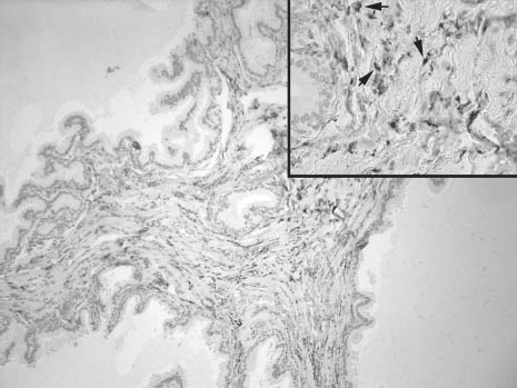
Immunohistochemical staining for PCV2 antigen was performed on selected formalin-fixed, paraffin-embedded tissues (lung, lymph nodes, tonsil, spleen, testis, seminal vesicles, prostate gland, bulbourethral glands, and intestine) using a rabbit polyclonal antiserum as previously described.10 Lung, lymphoid tissues, intestine, bulbourethral glands, epididymides, testes, and seminal vesicles were positive for PCV2 antigen. In the reproductive organs, PCV2-specific staining was observed in the cytoplasm of macrophages and fibroblast-like cells in the interstitium of all accessory sex glands and was particularly abundant in the interstitium of the seminal vesicles (Figure 1).
Figure 1: Immunohistochemical staining, using polyclonal anti-porcine circovirus type 2 (PCV2) antibodies, of the seminal vesicles of a boar naturally infected with PCV2, Mycoplasma hyopneumoniae, and Pasteurella multocida type D (x100). Inset: Higher magnification (x 200). Intense cytoplasmic staining was observed in interstitial macrophages and fibroblast-like cells (arrows).
|
Three doses of extended semen from an ejaculate collected from the boar the week prior to necropsy were tested for porcine circovirus type 1 (PCV1) and PCV2 and for porcine parvovirus (PPV)-specific nucleic acids by multiplex nested PCR.11 All three doses were negative for PCV1 and PPV. One of the three doses was positive for PCV2. Attempts to isolate PCV2 from this sample were unsuccessful.
Discussion
Recently, our group reported on the reproduction of PMWS and severe respiratory disease in 7-week-old pigs coinfected with PCV2 and M hyopneumoniae.12 In that experiment, M hyopneumoniae potentiated PCV2 infection in naive pigs. In the field, pigs are commonly exposed to PCV2 between 6 and 15 weeks of age.13 Boar studs are in many ways different from grow-finish operations. The health requirements for incoming pigs and the biosecurity level are more rigorous and the population sizes are much smaller. In this case, disease in the boar was likely due to initial exposure to PCV2 relatively late in life, at a time when the boar was concurrently infected with M hyopneumoniae and P multocida type D. The PCV2-associated lymphoid lesions in the boar were mild to moderate and appeared to be either in the early or resolving stages.
It was of interest that there was abundant staining for PCV2 antigen in the parenchyma of the seminal vesicles and that PCV2 nucleic acids were present in the semen of this boar, although no remarkable inflammation was observed in the reproductive tract. Although PCV2 nucleic acids have been demonstrated in semen of experimentally infected boars,6 this is, to our knowledge, the first report of PCV2 in the parenchyma of the secondary sex glands in a naturally infected boar. In a study investigating PCV2 in 98 one-year-old, randomly-selected, healthy boars from 49 herds, 13 of 98 whole semen samples were positive by PCR and 26 of 98 whole semen samples were positive by semi-nested PCR.14 In 11 of the 98 whole semen samples, PCV2 was isolated in cell culture. The same study also investigated prevalence of PCV2 in seminal fluid, nonsperm cells, and sperm heads, and detected the greatest amount of PCV2 DNA in the seminal fluid and nonsperm fraction.14 Kennedy et al (2000)15 demonstrated PCV2 antigen in infiltrating macrophages in the tunica albuginea, in interstitial macrophages and in germinal epithelial cells in the testes, and in infiltrating macrophages in the epididymides of boars 24 to 29 days after they had been coinfected with PCV2 and PPV as 3-day-old piglets.
During the investigation for potential causes of subfertile semen production in this boar stud, another possible explanation for decreased fertility was identified. Some types of latex gloves exert toxic effects on boar spermatozoa; therefore, vinyl or nitrile gloves are recommended for collection by the gloved-hand technique.16 Several boxes of latex gloves from one production lot were found in the collection area during the clinical investigation of this case. Samples from the boxes in use were submitted to the University of Pennsylvania Reference Andrology Laboratory for in vitro sperm toxicity testing. It was confirmed that direct exposure to the glove material was detrimental to boar sperm motility. Since nonspermicidal gloves were also used, and no records were kept of which boars were collected with which gloves, the contribution of this factor to the problem remains unclear.
Heat stress in boars may contribute to poor semen quality and productivity,17 but was not considered a significant influence in this case. Heat stress as a primary cause of poor semen quality was ruled out early in the clinical investigation, as the boars were initially affected relatively early in the season (May through July), the facility was relatively small and provided adequate cooling (mechanical ventilation with evaporative pads), and the affected boars were housed in crates in the center of the room surrounded by nonaffected boars.
This is the first report of PCV2 infection and concurrent bacterial infection in a clinically ill boar with poor fertility. These findings suggest that PCV2 was part of the disease complex that contributed to illness in this boar. Boars were not vaccinated for M hyopneumoniae either in isolation or in the main stud. Developing strategies to immunize naive boars against PCV2, M hyopneumoniae, or both prior to entry into studs should be considered when boars are likely to be concurrently exposed to PCV2, M hyopneumoniae, and other bacterial agents. Prospective studies are warranted to investigate a PCV2 and M hyopneumoniae co-infection model in mature boars to better understand the potential impact of this combination on reproductive performance and PCV2 transmission.
Implications
- PCV2-associated lung and lymphoid lesions may be present in young adult boars as well as in growing pigs.
- Enhancement of PCV2 infection by concurrent M hyopneumoniae infection may occur in adults as well as in growing pigs.
- Studies should be conducted to investigate, develop, and test the efficacy of strategies to immunize boars against PCV2 to minimize the risk of clinical disease and shedding of PCV2 in semen.
References
1. Allan GM, Ellis JA. Porcine circoviruses: a review. J Vet Diagn Invest. 2000;12:3-14.
*2. Sorden SD. Update on porcine circovirus and postweaning multisystemic wasting syndrome (PMWS). Swine Health Prod. 2000;8:133-136.
3. West KH, Bystrom JM, Wojnarowicz C, Shantz N, Jacobson M, Allan GM, Haines DM, Clark EG, Krakowka S, McNeilly F, Konoby C, Martin K, Ellis JA. Myocarditis and abortion associated with intra-uterine infection of sows with porcine circovirus 2. J Vet Diagn Invest. 1999;11:530-532.
4. O'Connor B, Gauvreau H, West K, Bogdan J, Ayroud M, Clark EG, Konoby C, Allan G, Ellis JA. Multiple porcine circovirus 2-associated abortions and reproductive failure in a multisite swine production unit. Can Vet J. 2001;42:551-553.
*5. Ladekjær-Mikkelsen AS, Nielsen J, Storgaard T, Bøtner A, Allan G, McNeilly F. Transplacental infection with PCV-2 associated with reproductive failure in a gilt [letter]. Vet Rec. 2001;148:759-760.
6. Larochelle R, Bielanski A, Müller P, Magar R. PCR detection and evidence of shedding of porcine circovirus type 2 in boar semen. J Clin Microbiol. 2000;12:4629-4632.
*7. Cameron RDA. Sexual development and semen production in boars. Pig News Inf. 1987;8:389-396.
8. Christopher-Hennings J, Nelson EA, Nelson JK, Hines RJ, Swenson SL, Hill HT, Zimmerman JJ, Katz JB, Yaeger MJ, Chase CCL, Benfield DA. Detection of porcine reproductive and respiratory syndrome virus in boar semen by PCR. J Clin Microbiol. 1995;33:1730-1734.
9. Mengeling WL, Vorwald AC, Lager KM, Brockmeier SL. Diagnosis of porcine reproductive and respiratory syndrome using infected alveolar macrophages collected from live pigs. Vet Microbiol. 1996;49:105-115.
10. Sorden SD, Harms PA, Nawagitgul P, Cavanaugh D, Paul PS. Development of a polyclonal-antibody-based immunohistochemical method for the detection of type 2 porcine circovirus in formalin-fixed, paraffin-embedded tissue. J Vet Diagn Invest. 1999;11:528-530.
11. Kim J, Chae C. Multiplex nested PCR compared with in situ hybridization for the differentiation of porcine circoviruses and porcine parvovirus from pigs with postweaning multisystemic wasting syndrome. Can J Vet Res. 2003;67:133-137.
12. Opriessnig T, Thacker EL, Yu S, Fenaux M, Meng XJ, Halbur PG. Experimental reproduction of postweaning multisystemic wasting syndrome in pigs by dual-infection with Mycoplasma hyopneumoniae and porcine circovirus type 2. Vet Pathol. 2004;41:624-640.
13. Larochelle R, Magar R, D'Allaire S. Comparative serologic and virologic study of commercial swine herds with and without postweaning multisystemic wasting syndrome. Can J Vet Res. 2003;67:114-120.
14. Kim J, Han DU, Choi C, Chae C. Simultaneously detection and differentiation between porcine circovirus and porcine parvovirus in boar semen by multiplex seminested polymerase chain reaction. J Vet Med Sci. 2003;65:741-744.
15. Kennedy S, Moffett D, McNeilly F, Meehan B, Ellis J, Krakowka S, Allan GM. Reproduction of lesions of postweaning multisystemic wasting syndrome by infection of conventional pigs with porcine circovirus type 2 alone or in combination with porcine parvovirus. J Comp Path. 2000;122:9-24.
16. Ko JCH, Evans LE, Althouse GC. Toxic effects of latex gloves on boar spermatozoa. Theriogenology. 1989;31:1159-1164.
*17. Blanc CD, Althouse GC. Seasonal infertility in an AI boar stud: a case report. Proc AASP. Nashville, Tennessee. 1996;27:163-166.
* Non-refereed references.


