| Case report | Peer reviewed |
Cite as: Caspari K, Henning H, Schaller C, et al. Semen quality and quantity in a boar with a complex hydrocele. J Swine Health Prod. 2012;20(4):174–178.
Also available as a PDF.
SummaryThis case report concerns a 3.5-year-old Duroc boar with an enlargement of the left hemiscrotum. Sonography revealed multiple hypoechogenic cavities around the testis, indicating a complex hydrocele. An infectious origin of the hydrocele was excluded by laboratory tests. Repeated semen analysis by microscope, computer-assisted sperm analyzer, flow cytometric assessment, and sperm chromatin structure assay showed only a subtle increase in proportion of sperm with head deformities 7 weeks after first clinical signs were observed. Necropsy confirmed the sonographic findings. The left testis was only half the size of the right. Histological examination of the left testis revealed moderate interstitial fibrosis, and areas of calcification were found. Signs of spermatogenesis were detected in only a few areas. Analysis of semen production data showed that the number of sperm per ejaculate was lower for this boar at the age of 2.5 to 3.5 years than at the age of 1.5 to 2.5 years (P < .001). To our knowledge, this is the first report of a complex hydrocele in a boar in the context of semen quantity and quality. | ResumenEste reporte de caso concierne a un macho Duroc de 3.5 años de edad con un agrandamiento del hemiescroto izquierdo. La sonografía reveló múltiples cavidades hipoecogénicas alrededor del testículo, indicando un hidrocele complejo. Se excluyó un origen infeccioso del hidrocele a través de pruebas de laboratorio. El análisis repetido del semen por medio del microscopio, analizador computarizado de esperma, evaluación mediante citometría de flujo, y prueba de estructura de cromatina de esperma mostraron solamente un incremento ligero en la proporción de esperma con deformidades de cabeza 7 semanas después de que se observaron los primeros signos clínicos. La necropsia confirmó los hallazgos sonográficos. El testículo izquierdo presentaba sólo la mitad del tamaño del derecho. El examen histológico del testículo izquierdo reveló fibrosis intersticial moderada, y se encontraron áreas de calcificación. Se detectaron signos de espermatogénesis sólo en algunas áreas. Los datos del análisis de la producción de semen mostraron que el número de esperma por eyaculación fue más bajo en este macho a los 2.5 a 3.5 años de edad que a los 1.5 a 2.5 años de dad (P < .001). Hasta donde sabemos, este es el primer reporte de un hidrocele complejo en un macho en el contexto de cantidad y calidad de semen. | ResuméCe rapport de cas intéresse un verrat Duroc âgé de 3.5 ans présentant une augmentation du volume de l’hémi-scrotum gauche. L’échographie a révélé la présence de multiples cavités hypo-échogènes autour du testicule, indiquant une hydrocèle complexe. L’origine infectieuse de l’hydrocèle a été exclue suite aux analyses de laboratoire. Les analyses répétées de la semence par microscopie, par analyseur de sperme assisté par ordinateur, par évaluation en cytométrie de flux, et par épreuve de la structure de la chromatine spermatique n’ont démontré qu’une faible augmentation de la proportion de spermatozoïdes avec des déformations de la tête 7 semaines après que les premiers signes cliniques furent observés. La nécropsie confirma les trouvailles échographiques. La taille du testicule gauche n’était que la moitié de celle du testicule droit. L’examen histologique du testicule gauche a révélé une fibrose interstitielle modérée, et des sites de calcification ont été trouvés. Des signes de spermatogénèse ont été détectés seulement dans quelques régions. L’analyse des données de production de semence a démontré que pour ce verrat le nombre de spermatozoïdes par éjaculat était inférieur à l’âge de 2.5 à 3.5 ans qu’à l’âge de 1.5 à 2.5 ans (P < .001). Selon nous, il s’agit de la première fois qu’un rapport fait état d’une hydrocèle complexe chez un verrat relativement à la quantité et la qualité de la semence. |
Keywords: swine, boar, hydrocele, sperm quality, sperm quantity
Search the AASV web site
for pages with similar keywords.
Received: November 3, 2011
Accepted: February 13, 2012
A hydrocele testis is a pathological accumulation of serous fluid between the layers of the tunica vaginalis that occurs when production of fluid by the vaginal tunic is increased or resorption is decreased.1 Hydroceles are described in several other domestic mammals and also in humans,2-7 appearing unilaterally or bilaterally as variable degrees of fluid enlargement of the scrotum without pain. They can be caused by scrotal trauma, infection, testicular neoplasia, or surgical procedures, or they may be idiopathic.1,8 Since the vaginal cavity communicates with the peritoneal cavity in most domestic mammals, hydroceles may form as an extension of ascites.9,10 The prevalence of hydrocele in infertile men was nearly twice as high as in a fertile control group.11 Dysfunction of spermatogenesis is thought to occur due to insufficient temperature regulation of the testis because of the accumulated fluid between the layers of the tunica vaginalis.1
Case history
Clinical signs and sonography
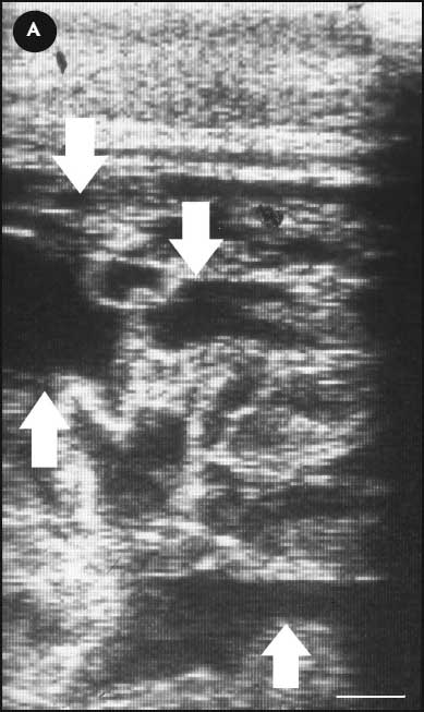
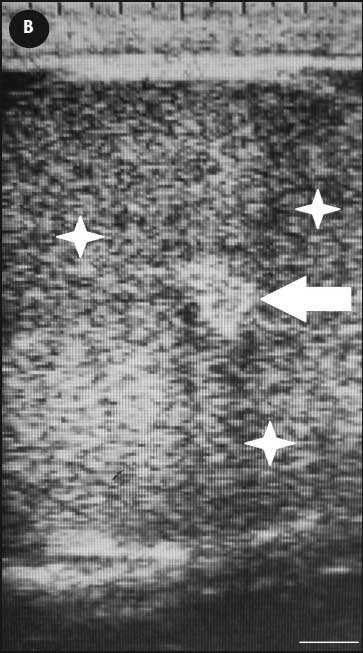
A Duroc boar was used for artificial insemination in a pig breeding unit. When the boar was 3.5 years old, an increasing enlargement of the left hemiscrotum was observed. The owner noted the first sign of enlargement approximately 4 weeks before the boar was examined by the veterinarian. Palpation revealed that the left hemiscrotum was enlarged approximately 2.5 times the size of the right, but remained painless. A fluctuant nondisplaceable mass surrounded the left testis, and gritty material was palpable between the scrotal skin and the testis. No other clinical signs were observed. Ultrasonography (Tringa Linear 5 MHz; Esaote Pie Medical, Cologne, Germany) of the left hemiscrotum revealed multiple hypoechoic cystic cavities (Figure 1A). The right hemiscrotum had a normal texture and no abnormalities (Figure 1B).12
Figure 1: Transcutaneous sonograms of the right and left hemiscrotums of a 3.5-year-old Duroc boar with a hydrocele testis of the left hemiscrotum. The linear transducer was placed frontally in the middle of the caudal part of the each hemiscrotum. (A) Left hemiscrotum: closest to the transducer are scrotal skin and tunica vaginalis lamina paritalis. Multiple hypoechogenic cystic cavities are seen within the processus vaginalis (white arrows). (B) Right hemiscrotum: closest to the transducer are scrotal skin and tunica vaginalis lamina paritalis et visceralis. The fine homogeneity of the testis is seen (white stars), with a hyperechogenic area of the rete testis (white arrow). (Bars = 1 cm).   |
Hematology and serology
Blood was collected from the superficial part of the genus descendens vein in the medial stifle area during semen collection twice at an interval of 3 weeks. Whole blood as well as blood in anticoagulant was collected each time. Samples were analyzed using an ST 2000iV hematology instrument (Sysmex Corporation, Japan). The hemogram showed normal to slightly increased values of unsegmented neutrophil granulocytes (1.1 and 3.0 × 10⁹ per L, respectively; reference range 0 to 1.5 × 10⁹ per L),13 segmented neutrophil granulocytes (11.1 and 4.2 × 10⁹ per L, respectively; reference range 1.0 to 8.2 × 10⁹ per L),13 and eosinophils (0.63 and 1.80 × 10⁹ per L, respectively; reference range 0 to 1.3 × 10⁹ per L).13
A rose bengal plate agglutination test was negative for Brucella suis antibodies. A microscopic agglutination test was negative for Leptospira interrogans serovars pomona, canicola, icterohaemorrhagiae, hardjo, bataviae, bratislava, autumnalis, australis, tarassovi, grippotyphosa, sejroe, and ballum.
Spermatology
Parallel to clinical investigation and approximately 1 week after the previous ejaculate had been collected, three ejaculates within 3 weeks were investigated in detail to assess the influence of the clinical findings with respect to semen quality. Morphology of 200 sperm cells was analyzed by phase-contrast microscopy at 1000× magnification (Axiostar plus; Carl Zeiss Feldbach, Germany) after fixation in a formol citrate solution.14 The first and third ejaculates were marginally above the reference value of 15.0% for retained cytoplasmic droplets (16.0%, 6.0%, and 15.5%, respectively).15 The first and second ejaculates each had 2.0% abnormal heads, within the normal range of a maximum of 5% abnormal heads. Only the third ejaculate contained an elevated number of sperm with deformed heads (8.0%; limit value 5.0%).15 Furthermore, the total number of morphologically abnormal sperm in the third ejaculate (30.0%) was above the reference range of ≤ 25.0%.15 Because the morphology of spermatozoa in the first ejaculate was essentially normal, the second and third ejaculates were evaluated in more detail. The motility of diluted spermatozoa was assessed by means of a computer-assisted sperm analyzer (Sperm Vision; Minitüb GmbH, Tiefenbach, Germany) 24 hours, 48 hours, and 72 hours after collection. Total motility in stored samples was within the reference range of ≥ 65% at all times.15 After 24 hours of storage, the proportions of sperm with defective plasma or acrosomal membranes or both were evaluated. Spermatozoa were stained with propidium iodide-conjugated peanut agglutinin (PI-PNA) and fluorescein isothiocyanate-conjugated peanut agglutinin (FITC-PNA) for analysis in a flow cytometer. The proportions of sperm with membrane defects (PI-PNA or FITC-PNA or both positive) in both samples were 20.8% and 10.8%, respectively (Table 1). Thus, both samples ranged below the proposed limit value of 26.5% membrane defects for diluted semen after 24 hours storage at 17°C.16 In addition, the numbers of sperm with unstable chromatin were evaluated with the sperm chromatin structure assay as previously described.17 A total of 2.8% and 2.0% of sperm per sample with a high DNA fragmentation index are in accordance with recently published mean values of 2.1%18 and 2.2%19 for semen doses stored for 24 hours. Both samples from the Duroc boar were below a proposed limit value of 5% for selection purposes as discussed by other authors.18,19 The results of the ejaculate analyses are presented in Table 1. The boar was euthanized because of the increase in number of sperm with abnormal head morphology.
Table 1: Results of analyses of three ejaculates collected weekly from a 3.5-year-old Duroc boar diagnosed with a hydrocele of the left hemiscrotum
CASA = computer assisted sperm analyzer; ND = not done; DFI = DNA fragmentation index. |
||||||||||||||||||||||||||||||||||||||||||||||||||||||||||||||||||||||||||||||||||||||||||||||||||||||||||||||||||||||||||||||||||||||||||||||||
Pathology
Necropsy examination revealed a gross thickening of the testicular envelope and fibrous pockets of brownish watery fluid between the parietal and visceral layers of the tunica vaginalis. The left testis was half as large as the right. There were no other macroscopic findings. Microscopically, the left testicular envelope showed severe fibrosis with perivascular infiltration of lymphocytes, macrophages containing hemosiderin, and small numbers of plasma cells. No neutrophil granulocytes could be seen. In the left testis there was moderate interstitial fibrosis with sporadic small areas of calcification. Only minimal spermatogenesis with matured spermatids could be detected in the tubuli seminiferi. No sperm were observed in the left epididymis. The right testis had normal structure with active spermatogenesis, and the right epididymis showed no lesions and harbored a moderate number of sperm cells.
Semen production history
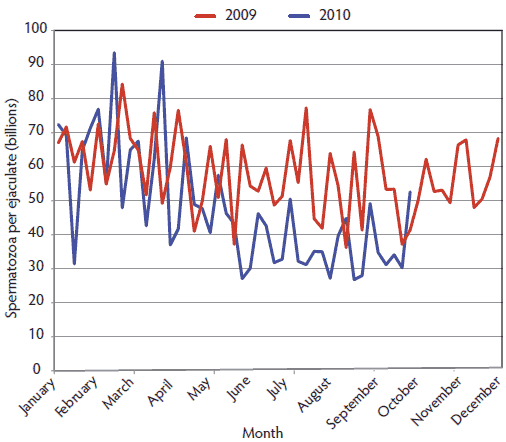
The sperm output of the boar during his productive phase was re-evaluated. Semen had been collected every 4 to 7 days for the previous 2.5 years. After each semen collection, the total number of spermatozoa per ejaculate was calculated from the volume and concentration of the raw semen. Volume was measured indirectly by electronic scale (Mettler; IMV-Technologies, L’Aigle, France) and concentration was evaluated by photometry (AccuCell; Boar Semen Photometer, IMV-Technologies). In 2009, the sperm output of the boar averaged 58.1 ± 11.4 billion sperm per ejaculate (n = 56). In 2010, the mean was 47.5 ± 17.3 billion sperm per ejaculate (n = 45) (Figure 2). This was a significant decrease in sperm output for this boar at the age of 2.5 to 3.5 years (2010) compared to the age of 1.5 to 2.5 years (2009) (P < .001; unpaired t test).
Figure 2: Total spermatozoa per ejaculate during the years 2009 and 2010 for a 3.5-year-old Duroc boar. A complex hydrocele of the left hemiscrotum was diagnosed by use of ultrasonography (2010) and confirmed at necropsy performed soon after diagnostic testing that showed an increase in numbers of sperm with abnormal head morphology.  |
Discussion
In the present case, the clinical appearance of the boar matched signs of a hydrocele as previously described by several authors.2-7 Ultrasound serves as an important imaging modality in all types of scrotal pathology.19 Simple hydroceles are anechoic or may contain low-level echoes secondary to fibrin bodies or cholesterol crystals. On the contrary, complex hydroceles may contain many internal echoes with septations,20 as in this case. Hydroceles may be caused by scrotal trauma, infection, testicular neoplasia, or surgical procedures, or they may be of idiopathic origin.1,8 No surgeries had been performed on this boar. Neoplasia as a cause of the hydrocele was excluded by pathological and histological examination. The septation observed in this hydrocele may occur as a consequence of infection or hemorrhage.21,22 Results of clinical examination, serology, and pathology gave no evidence of infection. Although normal to slightly increased numbers of white blood cells were observed in the blood samples, this was likely because blood was collected during semen collection. An increase of up to 40% of the reference value of white blood cells can be due to physical stress, eg, mounting a dummy.23 The pathological finding of hemosiderin in the macrophages indicates an insult (trauma) which could have caused hemorrhage. It is unknown if scrotal trauma with hemorrhage had occurred in this animal. Therefore, the cause of the complex hydrocele in this boar appeared to be traumatic but remains unclear. The total number of sperm per ejaculate was presumably affected by the hydrocele. Normally, sperm output tends to increase up to the age of 3.5 years.24 In this boar, the mean number of sperm per ejaculate had decreased significantly. The histopathological findings strengthen the assumption that a prolonged destructive process took place in the left testis. Proportion of damaged heads in the last collected ejaculate was increased. Damaged sperm heads are classified as a specific or nonspecific primary abnormality of the sperm cell, originating in the testis during spermatogenesis.25 Specific primary abnormalities are rare and seem to be congenital.25 Nonspecific primary abnormities are acquired and caused by an insult.25 Insufficient thermoregulation of the scrotum, along with other specific and nonspecific factors, may increase the proportion of abnormal sperm.26 Insufficient thermoregulation caused by the enlargement of the left hemiscrotum may explain the increased number of sperm with abnormal heads in this boar. It is not clear whether semen quality was affected before the hydrocele became clinically obvious. Until then, semen quality, except for motility, was not evaluated on a regular basis.
This case contributes to clinical findings in genital disorders of boars. This is, to our knowledge, the first report of a complex hydrocele in a boar in the context of semen quantity and quality.
Implications
• Decreasing sperm output can be an indicator of testicular disorders in boars.
• The testes of boars used for artificial insemination should be clinically observed and palpated on a regular basis to avoid unproductive days.
References
1. Schumacher J, Varner DD. Surgical correction of abnormalities affecting the reproductive organs of stallions. In: Youngquist RS, Threlfall WR, eds. Current Therapy in Large Animal Theriogenology. 2nd ed. St Louis, Missouri: Saunders Elsevier; 2007:26.
2. Patnaik AK, Liu S-K. Leiomyoma of the tunica vaginalis in a dog. Cornell Vet.1975;65:228–231.
3. Hopkins SM, Larsen RE, Drost M. Unilateral castration as treatment for hydrocele in a bull. JAVMA. 1981;178:837–838.
4. Fouad K. Hydrocele vaginalis testis in a stallion [in German]. Tierärztliche Praxis. 1980;8:479–480.
5. Brass KE. Die Sonographie in der andrologischen Untersuchung bei verschiedenen Haussäugetierarten [doctoral thesis]. Hanover, Germany: University of Veterinary Medicine Hanover; 1987.
6. Busch W. Andrology of the bull, pathology of the testis, the epididymis and the scrotum [in German]. In: Busch W, Holzmann A, eds. Veterinärmedizinische Andrologie, Stuttgart, Germany: Schattauer Verlag; 2001:199–212.
7. Uehara M, Takeda K, Tei H, Shimizu K, Imazu T, Yoshimura K, Kiyohara H. A case of liposarcoma of spermatic cord with a hydrocele. Acta Urologica Japonica. 2010;56:127–129.
8. Behre HM, Zitzmann M. Visual examination procedures [in German]. In: Nieschlag E, Behre HM, Nieschlag S, eds. Andrologie. 3rd ed. Heidelberg, Germany: Springer Verlag; 2009:108.
9. Ladd PW. The male genital system. In: Jubb KVF, Kennedy PC, Palmer N, eds. Pathology of Domestic Animals. 2nd ed. Orlando, Florida: Academic Press; 1985:409.
10. Abitt B, Fiske RA, Craig TM, Bitter JW. Scrotal hydrocele secondary to ascites in 28 bulls. JAVMA. 1995;207:753–756.
11. Qublan HS, Al-Okoor K, Al-Ghoweri AS, Abu-Qamar A. Sonographic spectrum of scrotal abnormalities in infertile men. J Clin Ultrasound. 2007;35:437–441.
12. Clark SG, Schaeffler DJ, Althouse GC. β-Mode ultrasonographic evaluation of paired testicular diameter of mature boars in relation to average total sperm numbers. Theriogenology. 2003;60:1011–1023.
13. Bickhardt K. Hämostase. In: Kraft W, Dürr UM, eds. Klinische Labordiagnostik in der Tiermedizin. 5th ed. Stuttgart, Germany: Schattauer Verlag; 1992:115–144.
14. Hancock JL. The morphology of boar spermatozoa. J Royal Microscopical Soc. 1956;76:84–97.
15. ZDS Guarantee requirements. Main association of German pig producers 2005 [in German]. Available at: http://www.zds-bonn.de/standardisierung.html. Accessed 10 April 2012.
16. Waberski D, Henning H, Petrunkina AM. Establishment of a sperm quality standard on pig AI stations. Reprod Dom Anim. 2010;45:55–56.
17. Evenson D, Jost L. Sperm chromatin structure assay is useful for fertility assessment. Methods Cell Sci. 2000;22:169–189.
18. Boe-Hansen GB, Christensen P, Vibjerg D, Nielsen MBF, Hedeboe AM. Sperm chromatin structure integrity in liquid stored boar semen and its relationship with field fertility. Theriogenology. 2008;69:728–736.
19. Waberski D, Schapmann E, Henning H, Riesenbeck A, Brandt H. Sperm chromatin structural integrity in normospermic boars is not related to semen storage and fertility after routine AI. Theriogenology. 2011;75:337–345.
20. Akin EA, Khati NJ, Hill MC. Ultrasound of the scrotum. Ultrasound Q. 2004;20:181–200.
21. Munden MM, Trautwein LM. Scrotal pathology in pediatrics with sonographic imaging. Curr Probl Diagn Radiol. 2000;29:185–205.
22. Stewart VR, Sidhu PS. The testis: the unusual, the rare and the bizarre. Clin Radiol. 2007;62:289–302.
23. Heinritzi K. Laboratory tests [in German]. In: Heinritzi K, Gindel HR, Reiner G, Schnurrbusch U, eds. Schweinekrankheiten. 1st ed. Stuttgart, Germany: Eugen Ulmer UTB; 2006:37–39.
24. Smital J. Effects influencing boar semen. Anim Reprod Sci. 2009;110:335–346.
25. Leidl W, Schefels W, Stolla R, Metzger E. Differentiation and fecundity of pathological forms of spermatozoa [in German]. Deutsche Tierarztliche Wochenschrift. 1971;78:129–134.
26. Weitze K-H. Examination of the spermatozoa [in German]. In: Busch W, Holzmann A, eds. Veterinärmedizinische Andrologie. Stuttgart, Germany: Schattauer Verlag; 2001;111–117.
