| Original research | Peer reviewed |
Cite as: Wattanaphansak S, Singer RS, Gebhart CJ. Evaluation of in vitro bactericidal activity of commercial disinfectants against Lawsonia intracellularis. J Swine Health Prod. 2010;18(1):11–17.
Also available as a PDF.
SummaryObjective: To evaluate the bactericidal activity of seven commercial disinfectants against Lawsonia intracellularis using a tissue culture system. Materials and methods: Two L intracellularis isolates were tested for susceptibility to several classes of disinfectants, including iodine, a biguanide, phenol, an oxidizing agent, a quaternary ammonium compound (QAC), and the combinations of QACs with aldehydes (formaldehyde and glutaraldehyde). All disinfectants were diluted with simulated hard water by adding 400 or 1000 ppm of calcium carbonate (CaCO3) and 5% fetal bovine serum (FBS). The effects of disinfectant concentrations (0.5×, 1×, and 2× the recommended dose) and exposure times (10, 30, and 60 minutes) at each concentration were investigated. Results: The susceptibilities of both L intracellularis isolates to the disinfectants were similar. When recommended doses of disinfectants were tested for 10 minutes at 400 ppm of CaCO3 and 5% FBS, both L intracellularis isolates were completely inactivated with QAC and the combinations of QAC with aldehydes. Inactivation was ≥ 99% with the oxidizing agent, 90% to 99% with the biguanide and phenol, and < 90% with iodine. At a CaCO3 concentration of 1000 ppm, the efficacies of combinations of QAC with glutaraldehyde were unchanged, while efficacies of the other disinfectants were reduced slightly. Implications: These data provide an in vitro guide for disinfectant selection to control L intracellularis. The results suggest that QAC, the combinations of QACs with aldehydes, and oxidizing agents would perform well for inactivation of L intracellularis under field conditions. | ResumenObjetivo: Evaluar la actividad bactericida de siete desinfectantes comerciales contra Lawsonia intracellularis utilizando un sistema de cultivo celular. Materiales y métodos: Se probaron dos aislamientos de L intracellularis en busca de la susceptibilidad a varias clases de desinfectantes, incluyendo yodo, un biguanide, fenol, un agente oxidante, un compuesto de cuaternarios de amonio (QAC por sus siglas en inglés), y las combinaciones de QACs con aldehídos (formaldehído y glutaraldehído). Todos los desinfectantes se diluyeron con agua dura simulada agregando 400 ó 1000 ppm de carbonato de calcio (CaCO3) y 5% de suero bovino fetal (FBS por sus siglas en inglés). Se investigaron los efectos de las concentraciones del desinfectante (0.5×, 1×, y 2× la dosis recomendada) y los tiempos de exposición (10, 30, y 60 minutos) en cada concentración. Resultados: La susceptibilidad de ambos aislamientos de L intracellularis a los desinfectantes fue similar. Cuando se probaron las dosis recomendadas de desinfectantes por 10 minutos a 400 ppm de CaCO3 y 5% FBS, ambos aislamientos de L intracellularis se inactivaron completamente con QAC y las combinaciones de QAC con aldehídos. La inactivación fue ≥ 99% con el agente oxidante, 90% a 99% con el biguanide y phenol, y < 90% con el yodo. En una concentración de 1000 ppm de CaCO3, la eficiencia de las combinaciones de QAC con glutaraldehído no cambió, mientras que la eficiencia de otros desinfectantes se redujo ligeramente. Implicaciones: Esta información provee una guía in vitro para la selección de desinfectantes para controlar la L intracellularis. Los resultados sugieren que el QAC, las combinaciones de QAC con aldehídos, y agentes oxidantes tendrían un buen desempeño de inactivación de L intracellularis bajo condiciones de campo. | ResuméObjectif: Évaluer l’activité bactéricide de sept désinfectants commerciaux contre Lawsonia intracellularis à l’aide d’un système de culture tissulaire. Matériels et méthodes: Deux isolats de L intracellularis ont été testés pour déterminer leur sensibilité à plusieurs classes de désinfectants, incluant l’iode, le biguanide, le phénol, un agent oxydant, un ammonium quaternaire (QAC), et les combinaisons de QACs avec des aldéhydes (formaldéhyde et glutéraldéhyde). Tous les désinfectants étaient dilués avec de l’eau dure simulée en ajoutant 400 ou 1000 ppm de carbonate de calcium (CaCO3) et 5% de sérum fÅ“tal de veau (FBS). Les effets des concentrations de désinfectant (0.5×, 1×, et 2× la dose recommandée) et les temps d’exposition (10, 30, et 60 minutes) à chaque concentration ont été étudiés. Résultats: Les sensibilités des deux isolats de L intracellularis aux désinfectants étaient similaires. Lorsque les doses recommandées de désinfectant étaient testées pour 10 minutes à 400 ppm de CaCO3 et 5% de FBS, les deux isolats de L intracellularis étaient complètement inactivés par le QAC et les combinaisons de QAC et des aldéhydes. L’inactivation était ≥ 99% avec l’agent oxydant, 90% à 99% avec le biguanide et le phénol, et < 90% avec l’iode. À une concentration de 100 ppm de CaCO3, l’efficacité des combinaisons de QAC avec le glutéraldéhyde était inchangée, alors que l’efficacité avec les autres désinfectants était légèrement diminuée. Implications: Ces résultats fournissement un guide in vitro pour la sélection des désinfectants visant à contrer L intracellularis. Les résultats suggèrent que QAC, les combinaisons de QAC avec des aldéhydes, et les agents oxydants seraient performants pour inactiver L intracellularis en condition de terrain. |
Keywords: swine, proliferative enteropathy,
disinfectants, Lawsonia intracellularis, ileitis
Search the AASV web site
for pages with similar keywords.
Received: August 14, 2009
Accepted: September 8, 2009
Lawsonia intracellularis is an important enteric pathogen responsible for causing proliferative enteropathy (PE) in pigs, horses, and other species. The disease is economically important in pigs and mainly affects growing-finishing pigs with a variety of clinical signs. The acute form of PE in pigs manifests itself as bloody diarrhea, often with sudden death, and occurs mainly in mature pigs. The chronic form is often found in younger growing pigs and is characterized by chronic diarrhea and poor growth rates. The subclinical form results in slow growth without diarrhea.1
The mechanism of disease spread in herds is not fully understood, though infection among pigs is mainly transmitted through a fecal-oral route. Infected pigs can continuously shed the organism through feces and be a source of infection for up to 10 weeks, and the number of bacteria shed has been estimated to be as high as 7 × 10⁸ L intracellularis per gram of feces.2 Shed bacteria can remain viable and infective in pig feces for 2 weeks.3 Therefore, the horizontal transmission of PE among new susceptible pigs can easily occur as a continuous infectious cycle via fecal material and fomites.
To control PE outbreaks and transmission in swine herds, proper disinfection of housing, pens, and equipment is important to reduce or eliminate the number of active L intracellularis in the environment. However, little information is available on the efficacy of various disinfectants against L intracellularis. This is largely because L intracellularis is an obligately intracellular bacterium that propagates itself only inside the host cell. Thus, it is difficult to perform primary bacterial isolation or find a reliable and reproducible intracellular assay to measure the efficacy of disinfectants against L intracellularis, as well as against other obligately intracellular organisms.
To date, only two in vitro studies report bactericidal activity of some disinfectants against L intracellularis. One uses a conventional tissue-culture method3 and the other a modified tissue-culture method as well as a direct-count method with specific fluorescent staining.4 In the latter modified tissue-culture method, several disinfectants over various concentrations and exposure times can be tested simultaneously. Since the results obtained from the modified tissue-culture method are similar to the direct-count method, either assay is appropriate for determining the activity of disinfectants against L intracellularis.4
The objective of this study was to use the modified tissue-culture method to evaluate the in vitro bactericidal activity of seven commercial disinfectants commonly used on swine farms. The in vitro conditions were conducted using simulated hard water produced by adding calcium carbonate (CaCO3) at two levels, and using fetal bovine serum as organic material. Furthermore, the morphology of L intracellularis after exposure to these disinfectants was also investigated.
Materials and methods
Microorganism preparations
Two L intracellularis isolates were used in this study: strains PHE/MN1–00 and NWumn05, obtained and isolated from affected pigs in the United States in 2000 and 2005, respectively. Both isolates were stored at -72°C until use. Each isolate was grown for three passages in murine fibroblast-like McCoy cells to allow recovery from the frozen stage and to obtain 100% confluence of viable bacteria in the cell cultures. The bacteria were grown and harvested as described earlier.5,6 Each isolate of L intracellularis was tested in replicate and each replicate of bacterial preparation was independently prepared from ten T-175 tissue-culture flasks. The final concentration of each preparation was measured using the direct-count method with immunostaining as described earlier.5,7
Disinfectants
Seven commercial disinfectants were selected to represent several classes of products commonly used in the swine industry. The commercial disinfectants, their main active ingredients, and the concentrations recommended for use are summarized in Table 1. To compare the bactericidal activity of the disinfectants, all tested disinfectants were prepared to final concentrations of 0.5×, 1×, and 2×, where × is the concentration in the manufacturer’s label instructions (Table 1).
Table 1: Disinfectants prepared according to label instructions and tested against Lawsonia intracellularis
* Roccal-D Plus (Pfizer Animal Health, Peapack, New Jersey); DC&R (Neogen, Lexington, Kentucky); Synergize (Preserve International, Reno, Nevada); Nolvasan S (Fort Dodge Laboratories, Fort Dodge, Iowa); Certi-Dine (Certified Safety Manufacturing Inc, Kansas City, Missouri); Virkon-S (Antec International, Sudbury, Suffolk, UK); and Tek-Trol (Bio-Tek Industries, Atlanta, Georgia). |
Test procedures
All disinfectants were diluted with two concentrations of simulated hard water created by adding CaCO3 to distilled water at either 400 ppm or 1000 ppm to test the influence of water hardness on their effectiveness. Each concentration of simulated hard water also contained 5% fetal bovine serum (FBS) to represent the presence of organic material. Both concentrations of simulated hard water were freshly prepared as described elsewhere.8 The final pH of the simulated hard water used in this study was adjusted to 7.6 to ensure growth of any viable L intracellularis present.
The working solutions of each disinfectant were freshly prepared to final concentrations of 0.5×, 1×, and 2×. Eight mL of each disinfectant concentration was aliquoted into a 15-mL polypropylene tube, and 300 μL of L intracellularis suspended in sterile phosphate buffered saline, containing approximately 108 L intracellularis per mL, was added to each tube and mixed. The bacterial suspensions were then filtered through 5-μm filters and transferred to three 2-mL microcentrifuge tubes. The tubes were incubated at room temperature (22°C to 25°C) for 10, 30, or 60 minutes. After incubation, the bacterial suspensions were centrifuged at 12,000g for 3 minutes. The pellets were washed and resuspended three times with 1.8 mL of the combination of Dulbecco’s Modified Eagles Medium (DMEM) and 30% FBS. After the final wash, pellets were resuspended with 1 mL of tissue-culture medium (DMEM with 20% FBS, 0.5% amphotericin B, and 1% L-glutamine) for tissue-culture inoculation. The controls used at each time point were live L intracellularis in simulated hard water without exposure to disinfectant, and dead L intracellularis, for which the bacteria were inactivated using isopropyl alcohol for 30 minutes.6 In this study, each concentration of the tested disinfectants was evaluated in duplicate, and both strains of L intracellularis were tested twice in 400 and 1000 ppm concentrations of simulated hard water.
The effectiveness of the disinfectants against L intracellularis was evaluated using bacterial viability after exposure as the indicator. The viability of L intracellularis was measured using a modified tissue-culture method in which the bacteria were grown in 96-well tissue-culture plates as previously described.4,7 Briefly, after treatment with disinfectant, 100 μL of bacterial suspension was transferred into each well of a 96-well tissue-culture plate to infect 2-day-old McCoy cells in quadruplicate cultures. After 24 hours of incubation and then every day for 3 consecutive days, the culture medium was changed. After 5 days of incubation in an atmosphere of 8.0% oxygen, 8.8% carbon dioxide, and 83.2% nitrogen, culture medium was removed from the infected plates and they were fixed with a 50:50 mixture of acetone:methanol. Fixed plates were then stained with the modified immunoperoxidase monolayer assay (IPMA) procedure using Lawsonia-specific rabbit polyclonal antibody as previously described,7 and the number of heavily infected cells (HIC) was assessed. A cell was considered to be heavily infected if the number of L intracellularis inside the cell exceeded 30 organisms.9 The number of HICs were counted, using the number of HICs as viability indicators and comparing the number of HICs in the disinfectant-treated cultures to the number in the disinfectant-free control cultures.
Scanning electron microscopy
To observe bacterial morphology after exposure to the disinfectants, one L intracellularis isolate, NWumn05, was exposed to a 1× concentration of each tested disinfectant for 10 minutes, as described above. The bacterial suspensions were then processed for examination as described previously4 using a scanning electron microscope (Hitachi S-3500N VP-SEM; Hitachi High Technologies America, Inc, Schaumburg, Illinois).
Data analysis
The HIC count for exposure time and disinfectant concentration was expressed as the percent by which bacterial concentration was reduced relative to the untreated control samples. The percentages of L intracellularis surviving for each exposure time, concentration, and disinfectant were averaged across the two individual tests.
Results
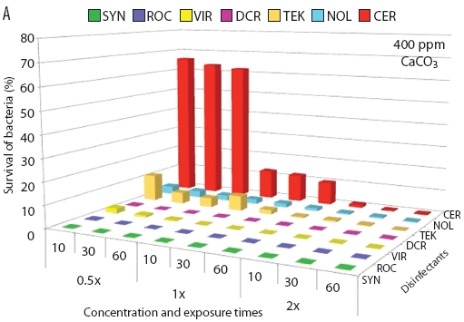
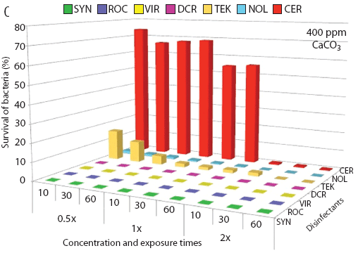
The in vitro susceptibilities of each L intracellularis isolate to the seven disinfectants are summarized in Figure 1. The results obtained from both L intracellularis isolates were similar with respect to susceptibility to disinfectants at the label-recommended concentrations (1×). When the disinfectants were diluted with water simulating 400 ppm of CaCO3 and 5% organic material, both L intracellularis isolates were completely inactivated within 10 minutes by QAC and the combinations of QAC with aldehydes (both formaldehyde and glutaraldehyde). Within 10 minutes of exposure, the oxidizing agent inactivated ≥ 99% of bacteria, phenol and biguanide inactivated 90% to 99% of bacteria, and iodine inactivated < 90% of bacteria. The surviving population of each L intracellularis isolate decreased as disinfectant exposure time increased from 10 minutes to 60 minutes and disinfectant concentration increased from 0.5× to 2× (Figures 1A and 1C).
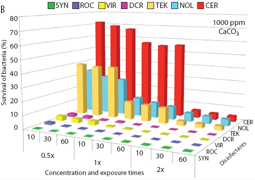
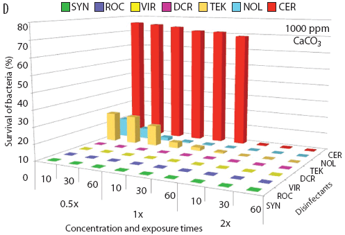
Figure 1: The effectiveness of seven disinfectants (described in Table 1) against Lawsonia intracellularis strains PHE/MN1-00 (A and B) and NWumn05 (C and D) in the presence of 5% fetal bovine serum (FBS) and CaCO3 at either 400 ppm (A and C) or 1000 ppm (B and D). The bacteria were exposed to 0.5×, 1×, or 2× the concentration of each disinfectant’s labeled dilution and incubated at room temperature for 10, 30, or 60 minutes before determining the bacterial viability using a modified tissue-culture method. Disinfectants included Synergize, a quaternary ammonium compound (QAC) with glutaraldehyde (SYN); Roccal-D Plus, a QAC (ROC); Virkon-S, an oxidizing agent (VIR); DC&R, a QAC with formaldehyde (DCR); Tek-Trol, a phenol (TEK); Nolvasan S, a biguanide (NOL); and Certi-Dine, an iodine (CER).
|
When the hardness of the synthetic water was increased to 1000 ppm of CaCO3, the efficacies of most of the tested disinfectants were reduced. However, the effectiveness of the QAC with glutaraldehyde combination was unchanged, and the QAC, the QAC with formaldehyde combination, and the oxidizing agent inactivated ≥ 99% of bacteria. The effectiveness of the remaining disinfectants decreased slightly more (Figures 1B and 1D).
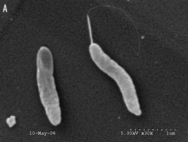
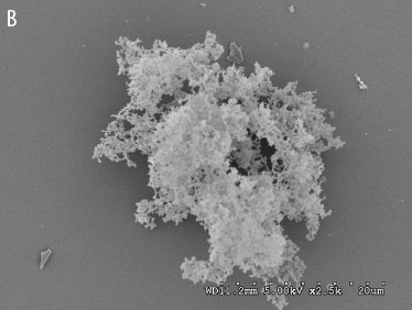
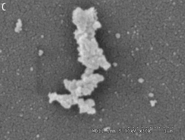
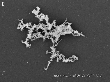
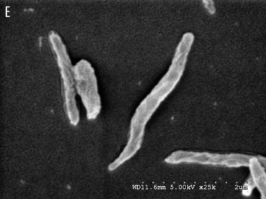
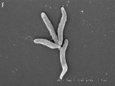
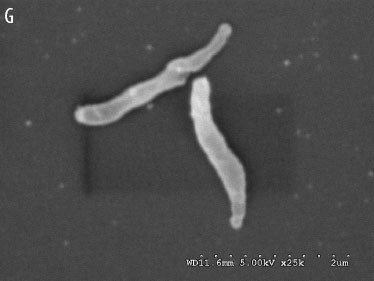
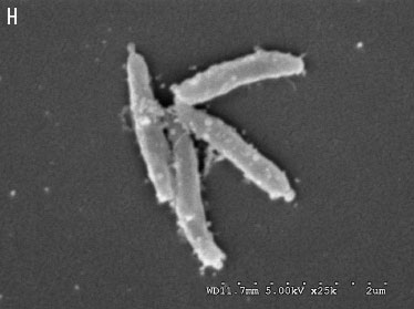
The morphology of L intracellularis was investigated after the bacteria were exposed to the recommended concentration of each disinfectant for 10 minutes. Scanning electron microscopy showed that most bacterial populations disappeared when treated with the QAC (Figure 2C), the combination of QAC with glutaraldehyde (Figure 2B), or the oxidizing agent (Figure 2D). Only the particle-like, ruptured L intracellularis cell membrane was found in each field of the electron microscope. In contrast, when bacteria were treated with the combination of QAC with formaldehyde (Figure 2E), the biguanide (Figure 2H), iodine (Figure 2D), or phenol (Figure 2F), the density of the bacterial population and the bacterial morphology, such as cell shape, size, flagellum, and membrane structure, were similar to those found in the control (Figure 2A).
Figure 2: Scanning electron microscopy showing the morphology of Lawsonia intracellularis strain NWumn05 after exposure to disinfectants (described in Table 1) for 10 minutes. (A) Lawsonia intracellularis was treated with phosphate buffered saline (control). Cells were exposed to (B) Synergize (a quaternary ammonium compound [QAC] with glutaraldehyde) at a concentration of 1:256, (C) Roccal-D Plus (a QAC) at a concentration of 1:256, (D) Virkon-S (an oxidizing agent) at a concentration of 1%, (E) DC&R (a QAC with formaldehyde) at a concentration of 1:128, (F) Tek-Trol (a phenol) at a concentration of 1:256, (G) Certi-Dine (an iodine) at a concentration of 1:128, and (H) Nolvasan S (a biguanide) at a concentration of 1:128.
|
Discussion
Although the mechanism of PE transmission among pigs and other animal species is not fully understood, it seems that bacterial contamination from feces and the environment play an important role. Antibiotics to which L intracellularis is susceptible eliminate the organism only from inside animals, while disinfectants inactivate L intracellularis only in the environment. Therefore, L intracellularis from both the animal and the environment should be eliminated and inactivated to reduce the potential for disease spread.
In the United States, water hardness levels vary across geographical regions and have been reported to range from 250 to 2848 ppm of CaCO3.10 Water hardness is considered very high when CaCO3 exceeds 300 ppm.10 More importantly, disinfectants are commonly used in environments containing organic materials such as feces, soil, and blood. Therefore, this study tested the disinfectants’ activities against L intracellularis using 5% FBS to mimic the presence of organic material. Of the disinfectants evaluated, QAC, the combinations of QAC with aldehydes (both formaldehyde and glutaraldehyde), and the oxidizing agent at the recommended concentrations were most active against L intracellularis in vitro.
The active ingredients of some tested disinfectants were a mixture of several compounds. It was beyond the scope of this study to determine the mechanisms by which the disinfectants inactivated L intracellularis. Morphological analysis of disinfectant-treated bacteria indicated particle-like debris from bacterial membranes after treatments with the QAC, the combination of QAC with glutaraldehyde, and the oxidizing agent. This suggests that these disinfectants might have the potential to lyse L intracellularis cells. The QAC and the combination of QAC with glutaraldehyde have membrane-active agents that cause cell-wall lysis by autolytic enzymes.11 Alternatively, the oxidizing agent is a combination of peroxygen compounds that presumably denature essential proteins and cell-wall structure.11 As in our study, El-Naggar et al12 found that the morphological structure of Escherichia coli exhibited lysation after exposure to 0.25% of an oxidizing agent (Virkon-S) for 15 minutes. However, no notable change in E coli morphology was observed upon treatment with 0.03% oxidizing agent (Virkon-S) for 60 minutes. In our study, the morphology of the formaldehyde-treated L intracellularis appeared intact, as with the control. These findings were similar to the report of El-Naggar et al,12 which found that formalin-treated E coli display morphologic characteristics similar to the control.
In a previous in vitro study, Collins et al3 reported that L intracellularis was highly inactivated by QAC and 1% povidone-iodine, while the bacteria tolerated 30 minutes of exposure to a 0.33% phenolic compound and hydrogen peroxide-peracetic acid at a concentration of 0.0005%. There were differences in strain responses to 1% potassium peroxymonosulfate and 0.001% sodium hypochlorite. Several factors contribute to the difficulty in making direct comparisons between this study and the study by Collins et al.3 Major differences between the two studies include strains of L intracellularis used (United States versus United Kingdom origin), bacterial concentrations (108 versus 104 L intracellularis per mL), testing concentrations, and exposure times. Furthermore, results from Collins et al3 were obtained without simulation of water hardness or organic-material load. However, there were notable similarities among the results. As in the previous study,3 both US L intracellularis isolates in this study were highly susceptible to QAC-based disinfectants and potassium peroxymonosulfate, and tolerated the phenolic mixtures. In contrast to the previous study, povidone-iodine-based disinfectants showed less effectiveness against L intracellularis. The lower bactericidal activity of povidone-iodine is mainly attributable to the effects of organic material in the hard water. However, when the concentration of povidone-iodine was increased to 2× (1:64 dilution), the percentages of viable L intracellularis were dramatically reduced. Moore and Payne13 reported that the effect of organic material decreases with higher iodine concentrations.
The authors are unaware of any in-depth investigations regarding the tolerance of L intracellularis to disinfectants. This warrants further investigation, since several studies have reported that bacteria resistant to disinfectants have the potential to develop resistance to some antibiotics.14,15 Russell14 and Thomas et al16 reported that the incorrect use of disinfectants, or their use with sub-lethal dosages, could possibly generate bacterial populations resistant to disinfectants.
Several investigations have reported similar dose-response relationships, where higher disinfectant concentrations required shorter exposure times to inactivate organisms.4,12,17 Although increasing the concentrations of disinfectants increased bactericidal activity, this over-application has several disadvantages, including expense, increased toxicity to animals and laborers, and corrosion of metallic material. The 10-minute exposure time was chosen for all disinfectants because it is the minimum exposure time that is recommended by most disinfectant manufacturers for activity to occur. Our results also showed that the bactericidal activity of the disinfectants increased with prolonged exposure time. Therefore, when disinfectants are used under field conditions, extending the exposure time to the floor or equipment surface may also increase the effectiveness of the products.
Uncertainty remains as to the ability of an in vitro laboratory test to predict a disinfectant’s bactericidal activity on a swine farm. Most disinfectant investigations, including this study, have tested bactericidal activity against microorganisms under suspension conditions. In reality, it is difficult to predict a disinfectant’s efficacy against sessile bacteria located on a dry dirty surface. Several investigations have reported that the bactericidal activities of disinfectants tested against bacteria on dry surfaces are lower than against bacteria in suspension, although bacteria were completely inactivated when tested under suspension.16,17 Because several factors are involved in the efficacy of disinfectants under field conditions (including organic load, temperature, and water hardness), it is possible that the in vitro results of this study may not accurately predict disinfectant effectiveness in swine barns. Further evaluation of these disinfectants on dry surfaces or under field conditions is needed.
Implications
- This investigation’s side-by-side comparison of the in vitro activities of disinfectants against L intracellularis could serve as a guide for disinfectant selection to control L intracellularis in the environment.
- QAC, the combinations of QAC with aldehydes (both formaldehyde and glutaraldehyde), and oxidizing agents at the recommended concentrations are likely to perform well to inactivate L intracellularis under field conditions.
Acknowledgements
This study was supported in part by a grant from Preserve International. The authors attest that the opinions and work contained herein accurately reflect their opinions and not necessarily those of Preserve International. The authors would like to thank Benjawan Wijarn and Molly Freese for excellent technical assistance.
References
1. Lawson GH, Gebhart CJ. Proliferative enteropathy. J Comp Pathol. 2000;122:77–100.
2. Smith SH, McOrist S. Development of persistent intestinal infection and excretion of Lawsonia intracellularis by piglets. Res Vet Sci. 1997;62:6–10.
3. Collins AM, Love RJ, Pozo J, Smith SH, McOrist S. Studies on the ex vivo survival of Lawsonia intracellularis. J Swine Health Prod. 2000;8:211–215.
4. Wattanaphansak S, Singer RS, Isaacson RE, Deen J, Gramm BR, Gebhart CJ. In vitro assessment of the effectiveness of powder disinfectant (Stalosan® F) against Lawsonia intracellularis using two different assays. Vet Microbiol. 2009;136:403–407.
5. Guedes RM, Gebhart CJ. Comparison of intestinal mucosa homogenate and pure culture of the homologous Lawsonia intracellularis isolate in reproducing proliferative enteropathy in swine. Vet Microbiol. 2003;93:159–166.
6. Wattanaphansak S, Gebhart C, Olin M, Deen J. Measurement of the viability of Lawsonia intracellularis. Can J Vet Res. 2005;69:265–271.
7. Wattanaphansak S, Singer RS, Gebhart CJ. In vitro antimicrobial activity against 10 North American and European Lawsonia intracellularis isolates. Vet Microbiol. 2009;134:305–310.
8. Association of Official Analytical Chemists. Disinfectants. In: Horowitz W, ed. Official Methods of Analysis. 13th ed. Washington DC: Association of Official Analytical Chemists; 1980:56–68.
9. McOrist S, Mackie RA, Lawson GH. Antimicrobial susceptibility of ileal symbiont intracellularis isolated from pigs with proliferative enteropathy. J Clin Microbiol. 1995;33:1314–1317.
10. American Water Works Association. Lime softening. In: Water Supply Operations: Water Treatment. 3rd ed. Denver, Colorado: American Water Works Association; 2003:313–361.
11. McDonnell G, Russell AD. Antiseptics and disinfectants: Activity, action, and resistance. Clin Microbiol Rev. 1999;12:147–179.
12. El-Naggar MY, Akeila MA, Turk HA, El-Ebady AA, Sahaly MZ. Evaluation of in vitro antibacterial activity of some disinfectants on Escherichia coli serotypes. J Gen Appl Microbiol. 2001;47:63–73.
13. Moore SL, Payne DN. Type of antimicrobial agents. In: Fraise AP, Lambert PA, Maillard JY, eds. Principles and Practice of Disinfection, Preservation, and Sterilization. 4th ed. Malden, Massachusetts: Blackwell Publishing; 2004:8–97.
14. Russell AD. Mechanisms of bacterial insusceptibility to biocides. Am J Infect Control. 2001;29:259–261.
15. Sidhu MS, Sorum H, Holck A. Resistance to quaternary ammonium compounds in food-related bacteria. Microb Drug Resist. 2002;8:393–399.
16. Thomas L, Russell AD, Maillard JY. Antimicrobial activity of chlorhexidine diacetate and benzalkonium chloride against Pseudomonas aeruginosa and its response to biocide residues. J Appl Microbiol. 2005;98:533–543.
17. Møretrø T, Vestby LK, Nesse LL, Storheim SE, Kotlarz K, Langsrud S. Evaluation of efficacy of disinfectants against Salmonella from the feed industry. J Appl Microbiol. 2009;106:1005–1012.

